Hans A. Bechtel
Committing SINS and Other Acts of Infrared
Advanced Light Source, Lawrence Berkeley National Laboratory, Berkeley, California USA
Infrared beamlines at synchrotron facilties worldwide take advantage of the spectrally broad and bright source characteristics of synchrotron radiation to perform label-free spectroscopic imaging of individual living cells, novel materials, and unique samples obtained from the bottom of the ocean to the depths of space. However, diffraction and the long wavelengths of infrared light have traditionally limited the spatial resolution of infrared techniques to the micron scale. Synchrotron infrared nano-spectroscopy (SINS) overcomes this limitation by combining the synchrotron infrared source characteristics with scattering type, scanning near-field optical microscopy (s-SNOM). By focusing and scattering synchrotron IR light off a metallic AFM tip in close proximity to the sample, the SINS technique dramatically improves the spatial resolution by several orders of magnitude to < 25 nm while still enabling sensitive vibrational spectroscopy spanning the entire mid- and far-infrared regions. The Advanced Light Source (ALS) at Lawrence Berkeley National Laboratory operates two infrared beamlines with SINS instruments that are available to general users. In this talk, I will describe the technical aspects of the technique and highlight several applications to soft and hard matter systems, including proteins, organic semi-conductors, catalysts, 2D materials, and phase-change materials [1-5].
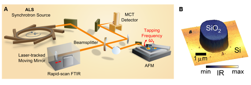
Figure 1. (A) Schematic of SINS experimental configuration. (B) SINS broadband IR signal response superimposed on 3D topography of an SiO2 microsctructure on silicon.
[1] H.A. Bechtel, E.A. Muller, R.L. Olmon, M.C. Martin, M.B. Raschke, PNAS 111, 7191–7196 (2014).
[2] E. A. Muller, B. Pollard, H. A. Bechtel, P. van Blerkom, M. B. Raschke. Science Advances, 2 (10), e1601006 (2016).
[3] C. Y. Wu, W. J. Wolf, Y. Levartovsky, H. A. Bechtel, M. C. Martin, F. D. Toste, Nature 541 (7638), 511 (2017).
[4] S. N. Gilbert Corder, et al. Nature Communications, 8, 2262 (2017).
[5] Z. Hao, H. A. Bechtel, T. Kneafsey, B. Gilbert, P. S. Nico, Scientific Reports, 8, 2552 (2018).
Dr. Carol Hirschmugl
Rapid 2D and 3D Infrared Imaging Applied to Biologically and Chemically Complex Systems
University of Wisconsin-Milwaukee, Department of Physics, Milwaukee, WI 53211 USA
*e-mail: cjhirsch@uwm.edu
The holy grail of chemical imaging is to provide spatially and temporally resolved information about heterogeneous samples on relevant scales. Synchrotron-based Fourier Transform infrared imaging1 combines rapid, non-destructive chemical detection with morphology at the micrometer scale, to provide value added results to standard analytical methods. Hyperspectral cubes of spatially and spectrally resolved data (x,y,z,Abs))) are obtained employing spectromicrotomography2, which is a label free approach. This method inherently evaluates a broad array of wide organic materials, with minimal sample preparation and modification. Examples presented here (polymer composites, single cells and colonies of cells) demonstrate the broad applicability of this approach to detect complex chemical information of intact samples.
References
1 Nasse, M. J., Walsh, M. J., Mattson, E. C., Reininger, R., Kajdacsy-Balla, A., Macias, V., Bhargava, R., and Hirschmugl, C. J. (2011) Nat.Methods 8, 413-416
2 Martin, M. C., Dabat-Blondeau, C., Unger, M., Sedlmair, J., Parkinson, D. Y., Bechtel, H. A., Illman, B., Castro, J. M., Keiluweit, M., Buschke, D., Ogle, B., Nasse, M. J., and Hirschmugl, C. J. (2013) Nat.Methods 10, 861-864
Acknowledgements This work was supported by the US NSF under awards CHE-0832298, CHE-1112433, and DMR-0619759, the Research Growth Initiative of the University of Wisconsin–Milwaukee, and is based on research conducted at the Synchrotron Radiation Center, University of Wisconsin–Madison which is supported by the University of Wisconsin– Milwaukee and University of Wisconsin–Madison. IRENI was supported in part by the Forest Products Laboratory and Forest Service.
Dr. Ferenc Borondics
Past, present and future of IR spectromicroscopy in SOLEIL
The SMIS infrared spectromicroscopy beamline provides unprecedented instrumentation that enables detailed investigation of scientific problems from biology to solid state physics. This is largely enabled by the instrumentation capability that allows six orders of magnitudes zoom-in on any sample, precise atmospheric, temperature and pressure control of the environment and the generation and detection of polarization. In this talk, I will review the currently available and upcoming instrumentation of the SMIS beamline through examples from user experiments. Finally, I will discuss an international collaboration led by the SMIS beamline that will revolutionalize data processing for spectroscopy.
Dr. Victoria Beltran
Discrimination of materials and its heterogeneities in stratigraphic samples from cultural heritage by µSR-FTIR
Victoria Beltran1, Balthazar Soulier2, Ferenc Borondics3, Alexandre Dazzi4, Christophe Sandt3, Ariane Deniset-Besseau4, Mathieu Thoury1, Loïc Bertrand1,3
1IPANEMA, CNRS MiC UVSQ, Université Paris-Saclay, BP48 Saint-Aubin, 91192 Gif-sur-Yvette cedex, France 2Atelier CELS, Paris, France 3Synchrotron SOLEIL, BP48 Saint-Aubin, 91192 Gif-sur-Yvette cedex, France 4Laboratoire de Chimie Physique, UMR 8000 CNRS, Université Paris-Sud, Université Paris-Saclay, 91405 Orsay, France
The application of organic coatings on historic artworks has been a constant practice throughout history. Coatings play an essential role to protect the pieces or to give them specific physical properties. The materials and techniques used in these layers are also a priceless testimony to understand the technology used by the artists and workshops [1]. The characterization of these coatings thus provides direct information not only about the most suitable conservation and restoration strategies but also about the historical background of each object.
However, the size of samples that can be studied, their stratigraphic structure, the wide range of compounds used and their alterations through time due to the interaction between themselves and with the atmosphere are strong constraints for their analysis [2]. FTIR proved its efficiency in discriminating between the main constituents of historical coatings and its alterations at the microscale [3]. Being a non-destructive technique, FTIR analysis can be integrated in multi-technique studies that allow to overcome the limits of a single technique. The ongoing development of high spatially resolved and high sensitivity imaging techniques allow the application of data processing that allow a finer depiction of such complex samples [4]. But such information is only attainable through a good analytical procedure which allows to go deep in the materials characterization and to understand its alteration processes.
A methodology coupling synchrotron and laboratory source-based analysis has been applied together with complementary techniques such as SEM-EDX to obtain the elemental composition and Optical Microscopy to improve the spatial resolution from the dozens of micrometers down to the nanometer. Challenges and limitations in terms of analysis and sample preparation will be discussed and illustrated with case studies including the results of the varnish analysis from a renaissance lute.
[1] De la Rie, E. R. (1989). Old master paintings: a study of the varnish problem. Analytical chemistry, 61(21), 1228A-1240A [2] Beltran, V., Salvadó, N., Butí, S., & Cinque, G. (2015). Micro infrared spectroscopy discrimination capability of compounds in complex matrices of thin layers in real sample coatings from artworks. Microchemical Journal, 118, 115-123. [3] Echard, J. P., & Bertrand, L. (2010). Complementary spectroscopic analyses of varnishes of historical musical instruments. Spectroscopy Europe, 22(2), 12. [4] Bertrand, L., Robinet, L., Cohen, S. X., Sandt, C., Le Hô, A. S., Soulier, B., … & Echard, J. P. (2011). Identification of the finishing technique of an early eighteenth century musical instrument using FTIR spectromicroscopy. Analytical and bioanalytical chemistry, 399(9), 3025-3032.
Raul Freitas (LNLS)
IR program at LNLS and future at SIRIUS
Synchrotron infrared (IR) spectroscopy is a long-established technique which explores the ultra-broadband IR spectral irradiance of electron storage rings for the chemical analysis of materials. This technique became available for the Brazilian scientific community in the last 4 years when the LNLS’ IR1 beamline became operational.
In this talk, I present the highlights of the IR program at LNLS and the perspectives for IMBUIA, the new IR beamline planned to operate in the new 4th generation accelerator SIRIUS.
Christiano de Matos (Mackgraphe)
Optical characterization of 2D and layered materials
MackGraphe – Graphene and Nanomaterials Research Center, Mackenzie Presbyterian University
Since the first isolation of graphene, optical methods have played a major role in the characterization of 2D materials and layered, van der Waals, crystals. The strong interaction of light with quasi-particles such as phonons and excitons also leads to the observation of polaritonic and nonlinear optical effects that may be of importance in future nanophotonic devices. In this talk, I will review some of the optical characterization activities carried out at MackGraphe. Through the optical characterization of black phosphorus, a layered semiconductor, for example, we have been able to identify edge phonons, strong optical nonlinearities mediated by light-exciton interaction and the early stages of oxidation. In the latter case, the synchrotron infrared nanospectroscopy system available at LNLS was used, enabling high spatial resolution and the spectroscopic identification of different oxide species.
Francisco Maia (LNLS)
Polaritons in Two-dimensional Materials and Heterostructures
Polaritons are, in general, electromagnetic modes coupled with fundamental resonances of matter. In two-dimensional (2D) crystals, the behavior of subdiffractional polaritons fully describes the nanophotonic properties. In this presentation, different types of polaritons (excitons-polaritons, plasmon-polaritons and phonon-polaritons) existing in different 2D materials, nanophotonic materials, will be presented. In special, an in-depth discussion will address to the hybrid photonic properties of heterostructures formed by the combination of graphene, hexagonal boron nitride (hBN) and talc. Either in graphene-hBN and graphene-talc, control of the key optical parameters (complex momenta, amplitude and phase of polaritonic waves) of midinfrared hybrid plasmon phonon-polaritons can be attained by an engineered architecture of nanodevices. It will be given a detailed description of scattering scanning near-field optical microscopy (s-SNOM) and nano-FTIR spectroscopy, which have being quasi-ubiquitous probes used in those studies. Since those techniques can directly interact and read the polaritons’ optical field, which happens in the near-field regime, they provide for reliable measurements. The advances in the knowledge of electromagnetism of polaritons and in the tools for measuring the optical near-field contribute to the development of the field of Nanophotonics. As an important prospect, such understandings can be useful for designing ultracompact devices with possible use in future nano-sensors, communication and computation mechanisms.
Thiago Miguel (LNLS)
Sample preparation strategies for micro and nano-FTIR
Sample preparation is a key step in the pipeline of advanced imaging modalities such as synchrotron nano-FTIR and micro-FTIR. In this talk, I present the most important steps in sample preparation for infrared analysis in the micro and nano-scales. In phase with the sample requirements for those techniques, which will be available in the new IR beamline at SIRIUS (IMBUIA endstation), I will present the plans for the supporting labs that will assist the users community on the sample preparation as an additional service connected to approved beamtime proposals.
Ingrid Barcelos (LNLS)
Fabrication Process, Challenges and Applications in New Two-Dimensional materials Beyond Graphene
Graphene’s success has shown that it is possible to create stable, single and few layers materials, and that these materials can exhibit fascinating and technologically useful properties. In the mid-infrared, graphene-based heterostructures and hexagonal boron nitride (hBN) overwhelmingly concentrate the attention by exhibiting real-space nano-optics features from plasmons, phonon−polaritons, and hybrid plasmon phonon− polaritons. Here, we show interesting optical properties of new 2D materials beyond graphene using Infrared Nano-spectroscopy. Initially, we will outline the different class of 2D materials and discuss the strategies to prepare single, multilayers and heterostructures. Moreover, we describe the infrared fingerprint of some these materials and other polaritonic properties. Additionally, we present the development of a robust fluidic chip use graphene as an optical window, whose contact area is then accessible to the tip-enhanced infrared nanoprobes, specifically designed for Infrared spectroscopy of liquid environments. We demonstrate the feasibility for accessing liquids and biological structures, as proteins. Finally, we highlight the advantages and challenges of 2D materials as a polaritonic platform, in optoelectronics devices, among other applications.
Ana Flavia Nogueira (Unicamp)
Synchrotron radiation applied to the characterization of perovskite films: morphology, structure and composition
Rodrigo Szostak1,2, Raul O. Freitas2, Helio C. N. Tolentino2, Ana Flavia Nogueira1
1University of Campinas (UNICAMP), Laboratório de Nanotecnologia e Energia Solar, Chemistry Institute, Campinas, PO Box 6154, 13083-970, Brazil
2Brazilian Synchrotron Light Laboratory (LNLS), Brazilian Center for Research in Energy and Materials (CNPEM), Campinas, SP, 13083-970, Brazil.
Organic inorganic hybrid perovskites (OIHP) is the most promising material to achieved high power conversion efficiency (PCE) at low cost. The high-quality optoelectronic properties in combination with solution-based preparation methods are responsible for the currently certified PCE record of 23.3%1, which is close to the PCE of monocrystalline silicon solar cells (26.1%). OIHP is generally labelled as an ABX3 compound, where A is a monovalent cation such methylammonium (MA), formamidinium (FA), or cesium (Cs), B is a divalent metal, such lead (Pb) or tin (Sn), and X is a halide anion, bromide (Br), iodide (I–). The properties of the perovskite film are direct related to film morphology, composition and crystalline structure, thus a clear understanding of how and when the intermediate and the perovskite phases are forming, as well the distribution of these multiple phases in the bulk and grains boundaries are important questions to be addressed in order to improve perovskite film properties and consequently the PCE of the devices. In this presentation we will summarize our most recent results using in situ time-resolved grazing incidence wide angle x-ray scattering (GIWAXS) and synchrotron infrared nanospectroscopy (nano-FTIR). GIWAXS experiments allowed us to understand the influence of the relative humidity, type of solvent and time to drop the antisolvent during the preparation of mixed cation perovskite films. We also identified intermediates formed during the spin coating process. Nano-FTIR technique was applied for the first time on OIHP. Our results revealed a spatial heterogeneity of the vibrational signal, which are associated to different chemical composition. The nano-FTIR permitted to access grain-to-grain chemistry of OIHP and the identification of PbI2 and hexagonal phases which are distributed randomly in a background formed by cubic (black phase) perovskite.
1.National Renewable energy Laboratory. Best Research Cell Efficiencies. Available at: https://www.nrel.gov/pv/assets/images/efficiency-chart.png.