Description
Nanomaterials have revolutionized the way we live in this world. The nanotechnology has enabled to tailor the structure of the material at an extremely small scale to achieve specific properties which have allowed to improve technology and industry sectors. The so-called Engineered Nanomaterials (ENMs) have produced many functional and innovative materials that are widely used in several applications, for instance, in cosmetics, medicine, energy, agriculture, environment, etc.., to name few of them.
Significant advances have been done in the past few years on studying the relationship between physicochemical properties of the nanomaterials and their toxicity. As for the environment, general concerns have raised about the effects on the ecosystems due to the extensive transformation of the ENMs, many of them with unknown properties. Studies in-vitro and in-vivo conditions should be conducted to properly understand their physicochemical properties and thus to assess the impact of these materials in complex systems. Among widely used traditional techniques for ENMs characterization, synchrotron radiation techniques have proved to be very useful as it can be possible to study these complex systems in a non-destructive manner offering high sensitivity, chemical specificity, and spatial resolution.
The main goal of this workshop is to discuss the status of the applications of synchrotron-based techniques to characterize ENMs in complex biological and environmental systems. Moreover, with the new Brazilian fourth generation light source Sirius entering soon in operation, synchrotron light with unprecedented intensity and brilliance will be delivered to the experimental stations allowing to perform novel and challenging experiments within the main topics addressed in this workshop.
Local: Plenary Room
Program
Invited Speakers Abstracts
 Short bio: Prof. Chunying Chen received her PhD degree in Biomedical Engineering from Huazhong University of Science and Technology of China in 1996. She joined the CAS Key Laboratory for Biomedical Effects of Nanomaterials & Nanosafety. Her research focuses on the Nano-Bio interactions, transformation and fate of nanomaterials in biological systems and therapies for malignant tumors using theranostic nanomedicine systems. She has been awarded the Second Prize of the National Natural Science Award in 2018, Outstanding Female Awards of the Chinese Academy of Sciences in 2017, and Chinese Young Female Scientists Award in 2014 and supported by the National Science Foundation for Distinguished Young Scholars of China. See more here.
Title: “Characterization of the pharamcokinetics and metabolites of metal-based nanomaterials in biological system”
Abstract: Nanomaterials possess high surface reactivity towards the biological matter, therefore, how to understanding nano-bio interactions of nanomaterials in the context of in vivo and the biological environments is crucial for the success of nanomaterials. In this talk, we will overview the developments of techniques and methods that allow reproducible and reliable analysis for absorption, distribution, metabolism and excretion (ADME) and biochemical characteristics of nanomaterials in vivo. ADME should also be capable of answering several key fundamental questions, i) where and how much nanomaterials get in? ii) where and how much NMs/NPs go? iii) how much, when and what form of nanomaterials remain intact? and iv) where, how much and what form of nanomaterials stay in the system?
We will discuss the analytical methodology and techniques developed for the quantification of the ADME processes of nanomaterials in vivo, focusing on those used for nanomaterials quantification in different bio-matrices, such as blood, tissues, organs and biomedical processes. To understand the full picture of nanomaterials metabolism in the liver, it is crucial to monitor in situ the dynamic changes in their concentration, composition and precise location with time, and to accurately quantify the metabolite products. Chemical speciation analytical techniques is highly necessary to study the chemical process and fates of nanomaterials, such as X-ray absorption spectroscopy (XAS) and high performance liquid chromatography (HPLC)-coupled ICP-MS (HPLC-ICP-MS), which can sensitively reveal the chemical structures and states of nanomaterials in the liver.
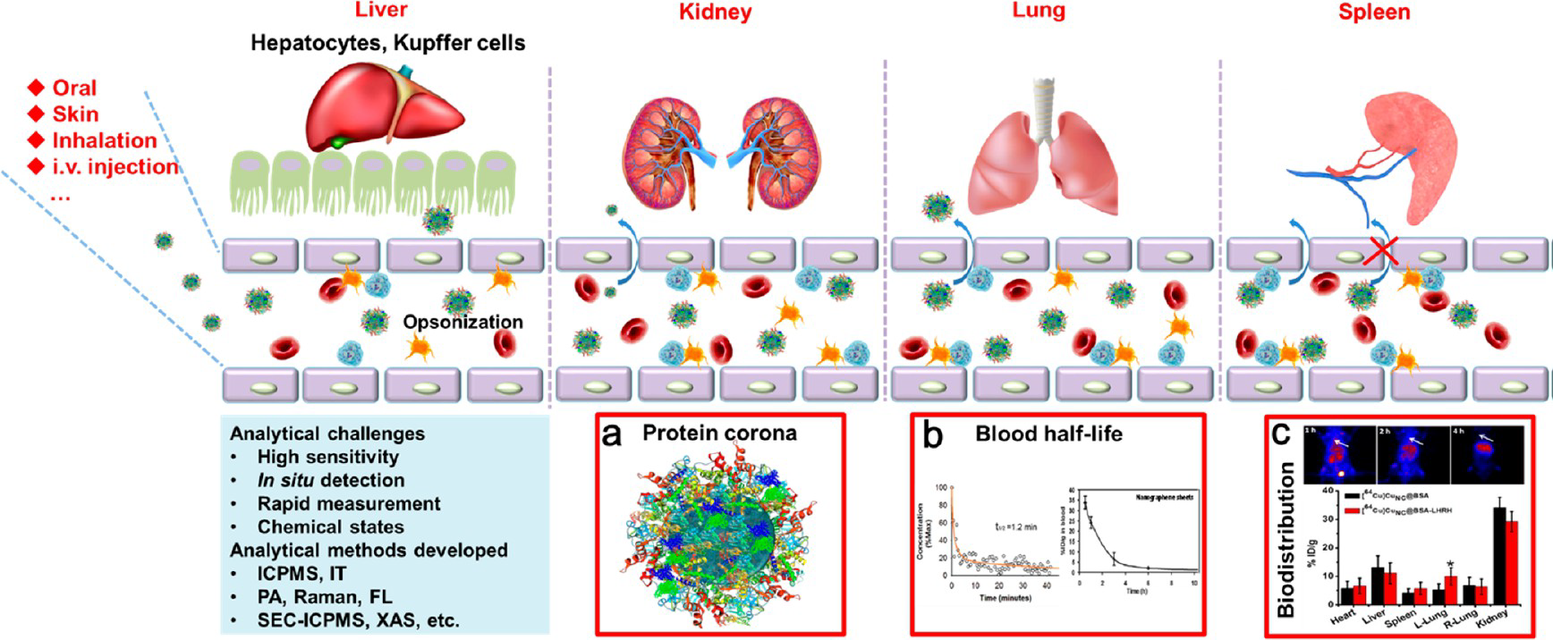 Figure 1 –Quantitative Analysis of Nanomaterials in vivo.
REFERENCES
- Wang, X.; Wang, M.; Chen, C.; et al. Chiral Surface of Nanoparticles Determines the Orientation of Adsorbed Transferrin and its Interaction with Receptors. ACS Nano, 2017, 11 (5), pp 4606–4616.
- Wang, X.; Wang, X.; Wang, M.; Chen, C.; et al. Probing Adsorption Behaviors of BSA onto the Chiral Surfaces of Nanoparticles. Small, 2018, 10.1002/smll.201703982.
- Chen, C., Wang, H. Biomedical Applications and Toxicology of Carbon Nanomaterials. 2016, John Wiley & Sons, Inc
- Wang, L., Zhang, T., Li, P.,, Chen, C., et al. Use of Synchrotron Radiation-Analytical Techniques to Reveal Chemical Origin of Silver-Nanoparticle Cytotoxicity. ACS Nano, 2015, 9(6): 6532-6547.
- Zhang, Z., Wang, J., Nie, X., Wen, T., Ji, Y., Wu, X., et al. Near Infrared Laser Induced Targeted Cancer Therapy Using Thermo-Responsive Polymer Encapsulated Gold Nanorods. J Am Chem Soc, 2014; 136:7317-7326.
- Wang, L.M., Li, J.Y., Pan, J., Jiang, X.M., Ji, Y.L., Li, Y.F., Qu, Y., Zhao, Y.L., Wu, X.C.*, Chen, C.Y.* Revealing the binding structure of the protein corona on gold nanorods using synchrotron radiation-based techniques: Understanding the reduced damage in cell membranes. J. Am. Chem. Soc., 135(46)(2013), 17359-17368.
- Chen, Z., Liu, Y., Sun, B., Li, H, Dong, J., Zhang, L., Wang, L., Wang, P., Zhao, Y., Chen, C. Polyhydroxylated Metallofullerenols Stimulate IL-1 Secretion of Macrophage through TLRs/MyD88/NF-κB Pathway and NLRP3 Inflammasome Activation. Small, 2014, 10(12): 2362-2372.
- Chen, C., Li, Y., Qu, Y., Chai, Z., Zhao ,Y. Advanced nuclear analytical and related techniques for the growing challenges in nanotoxicology. Chemical Society Reviews, 2013, 42(21):8266-303.
Title: Hyperspectral chemical imaging in space and time using synchrotron light.
Abstract: Commonly in technological functional/engineered materials as well as in nearly all inorganic or biological materials in nature, pronounced physical and chemical heterogeneities are
observed at various length and time scales. These complex hierarchical structures potentially dictate the macroscopic physical properties and chemical reaction pathways in space and time. Consequently, being able to unveil physicochemical properties at relevant spatial and temporal length scales is fundamental for the understanding of properties, function, and reactivity of materials.
Imaging is frequently limited to morphological analysis; however, very often imaging disclosing physicochemical properties turns out to be essential. Therefore, the need for “chemical microscopes” is growing rapidly. In that regard, synchrotron-based techniques have been applied to provide, in a non-destructive manner, high-resolution imaging of chemical speciation for various complexes and heterogeneous systems. The use of several Xray techniques providing chemical contrast such as X-ray fluorescence (XRF), X-ray absorption spectroscopy (XAS) or X-ray diffraction (XRD) in a simultaneous and/or combined manner allow the recording of multimodal and multidimensional/hyperspectral datasets. Among their advantages, it can be mentioned:(i) the potential to perform in-situ experiments;
(ii) resolution that can be achieved down to the tens of nanometers;
(iii) in addition to 2D scanning analysis studies (‘chemical images’) based on spectromicroscopy and XRD-imaging also, due to the considerable penetration power of (hard) xrays, investigations in 3D (‘chemical tomography’) are also feasible, which provides means to derive local chemical information from within intact, undisturbed objects or materials;
(iv) ability to get depth resolved crystalline contrast by analyzing the XRD and chemical–elemental contrast by analyzing the XRF;
(v) element-specific chemical sensitivity by tuning the X-ray energies around elementspecific absorption resonances.
In this presentation, recent progress and achievements in the field of 2D/3D chemical imaging and speciation analysis using various synchrotron radiation x-ray microprobe techniques, as well as on full-field ones will be presented. Examples from a wide range of scientific disciplines, including materials science, environmental science, or biology, will be presented, which will include applied research in to health and environmental sciences [1,2], energy [3–5] and catalysis [6–8]. Future prospects will be addressed and arising new research opportunities will be outlined.
1. Kapishnikov, S. et al.. Sci. Rep. 7, 1-12 (2017).
2. Sanchez, D. F. et al.. Sci. Rep. 7, 1-13 (2017).
3. Laurencin, J. et al.. Electrochim. Acta 241, 459-476 (2017).
4. Sanchez, D. F., Grolimund, D., Hubert, M., Bleuet, P. & Laurencin, J.. Int. J. Hydrogen Energy 1-9 (2016). doi:10.1016/j.ijhydene.2016.11.094
5. Tsai, E. H. R. et al.. iScience 11, 356-365 (2019).
6. Ihli, J. et al.. Angew. Chemie – Int. Ed. 56, (2017).
7. Ihli, J. et al.. Nat. Commun. 8, (2017).
8. Ranocchiari, M., Ferreira Sanchez, D. & Van Bokhoven, J. A. X-ray chemical nano-imaging to monitor chemical and structural changes in metal-organic framework HKUST-1. Prep.
 Bio: Acess here.
Title: “Sub-micron X-ray microspectroscopy for life and environmental nanotoxicology”
Abstract: Roughly 2000 products currently available on the market contain engineered nanomaterials (ENMS), and this number is increasing. The physical and chemical properties of ENMs differ from their bulk counterparts and their industrial applications range from electronics and optics to medicine and foods. Hence, understanding the fate, and physical and chemical modifications of ENMs in living organisms, to assess their positive or negative impacts, requires specialized analytical techniques. Since both chemical and physical factors must be considered for a better understanding of ENMs behaviour in complex matrices, these materials might be considered a new type of analyte. An ideal technique should offer the best balance between sensitivity, chemical specificity, and spatial resolution. In this sense, microspectroscopic techniques, as available at ESRF beamline ID21, are of special interest in life and environmental nanotoxicology.
Localization and speciation of elements at ID21 is done using micro-X-ray fluorescence (µXRF) and micro X-ray absorption spectroscopy (µ-XANES) in the tender X-ray domain (2-9.1 keV). The X-ray beam is focused using KB optics to a sub-micron spot (~500 nm), which then allows localization of trace elements at subcellular level. The beamline is equipped with a passively cooled cryogenic stage that allows the study of frozen hydrated specimens (cells and cryo-sectioned tissues) preventing elemental redistribution and minimizing radiation damage. The combination of µXRF and µXANES allows precise localization and speciation of biologically relevant (P, S, Cl, Ca, K) and widely used elements in nanotechnology (Cd, Ag, Ti, La, Ce) in complex samples.
This presentation will highlight research done at ID21 taking full advantage of the beamline capabilities to investigate the distribution and speciation of ENMs in complex environmental and biological samples (e.g. soils, tissues, and cells). In the context of EBS and ID21’s refurbishment project the main technical challenges and keystone developments to consider for the design of the new end-station will be discussed.
Title: Nanoparticles-cell interaction probed by x-ray microscopy
Abstract: The understanding of the mechanisms behind the interaction of nanoparticles and living cells is the cornerstone for the development of future nanomedicine. Improvements on nanoparticles efficiency and their safety regarding applications on diagnostics and therapeutics can fundamentally be traced back to the nanoparticle distribution within a cell. However, probing nanoparticles internalization and trafficking into cells with subcellular resolution remains limited by conventional 2D imaging or low-resolution 3D information. At the same time, the recent development of x-ray microscopes has pushed x-ray 3D imaging down to few nanometers in resolution. In this talk, I will show our first results where we could track nanoparticles inside cells with resolution better than 20 nm. Also, I will discuss the perspectives of studying nanoparticles-cells systems using Ptychographic Tomography and how it will allow the high-resolution imaging of multiple nanoparticles inside cells unveiling the fundaments behind both nanoparticle uptake and long-term exposure to advanced nanomedicines.
This is a satellite event to the 29th edition of the LNLS Annual Users Meeting (RAU).