SCIENCE HIGHLIGHTS

Cancer, ecological foam, machine learning, enzymes and fungi were just some of the topics of prominent articles published by CNPEM researchers between 2020 and 2021.

CNPEM’s internal research focuses on topics of interest to Brazil in sectors such as health, energy, new materials, the environment, and quantum technologies. The projects are influential and reliable knowledge bases for the national and international scientific community. Studies conducted at the Center have its state-of-the-art infrastructure at their disposal, along with essential and experienced specialists working in a multidisciplinary environment who are open to disruptive and visionary challenges.

Funding Agencies:

FAPESP – São Paulo Research & Innovation Support Foundation

CAPES – Brazilian Coordination for Development of Higher Education Personnel

CNPq – Brazilian National Council for Scientific and Technological Development

INCT-INOMAT – National Institute of Science and Technology in Complex Functional Materials

INCTBio – National Institute of Science and Technology for Bioanalytics

VIRUSES

The unique anatomy of the Mayaro virus

THE FIRST VIRAL STRUCTURE DOCUMENTED IN AN ENTIRELY BRAZILIAN EFFORT DESCRIBES THE UNIQUE AND FUNDAMENTAL CHARACTERISTICS OF THE MAYARO VIRUS. THIS RESEARCH WILL FACILITATE THE DEVELOPMENT OF NEW DIAGNOSTIC METHODS, MEDICATIONS, AND IMMUNIZATIONS AGAINST YET ANOTHER MOSQUITO-BORNE DISEASE.

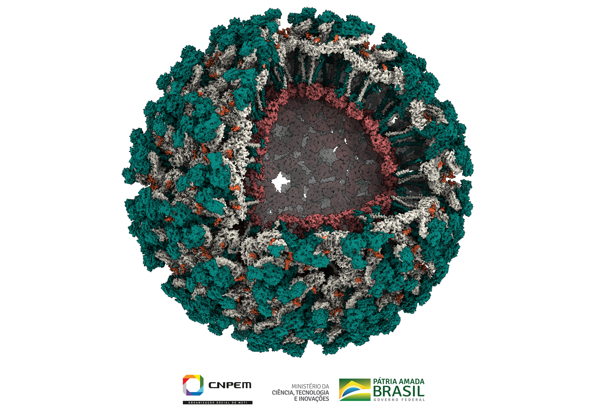
This is the first time that the complete structure of a virus has been described in Brazil and Latin America; this was accomplished by a multidisciplinary team of researchers from CNPEM.

The project, which was published in the journal Nature Communications, revealed unprecedented details of the molecular structure of the Mayaro virus at 4.4 angstrom resolution, approximately 100,000 times smaller than the thickness of a human hair.

The virus, which was first identified in Trinidad and Tobago in the 1950s, spread throughout the Americas. The infectious mosquito-borne illness known as Mayaro virus disease causes joint pain that can last for months. This is one of Brazil’s neglected endemic diseases; it is difficult to diagnose and has symptoms very similar to those of Chikungunya virus, which is a major hindrance to control strategies.

This project involved three and a half years of work by 20 researchers and collaborators, sophisticated electronic cryomicroscopy equipment, and advanced biology techniques to reveal the structure of the virus.

“In this project we described the infectious particle of the Mayaro virus, including all of its constituent proteins. We used techniques that allowed us to observe details of the biology of the virus that had not been described elsewhere, which represents an advance in our abilities to combat and understand this disease,” explains Rafael Elias Marques, a researcher at CNPEM.

One of the main techniques used to reveal the viral structure was electronic cryomicroscopy. This method, which makes it possible to see the three-dimensional atomic structure of biological molecules, was the focus of the 2017 Nobel Prize in Chemistry and has been widely used to understand details of other viruses, such as SARS-CoV-2.

“At CNPEM we run an electron microscopy center that is unique in Latin America. This set of equipment is open for the scientific community to use free of charge, and has been established to make Brazilian research in structural biology internationally competitive. This project is an outstanding example of this potential,” explains CNPEM researcher Rodrigo Portugal.

Cutaway representation of the Mayaro virus. Each of the proteins that comprise the virus particle is shown in a different color (green, gray, and red); sugars bound to the proteins are shown in orange.

HANDSHAKE

Notable in the newly revealed structure are the chains of sugars linked in the E2 protein. These sugars face each other in a configuration described as a handshake. Researchers believe that these sugars are recognized by the immune system, and can also help the virus organize and become more stable. The function of this specific part of the virus is among the topics that the researchers will continue to study.

“When we learn the details of the proteins that make up the structure of a virus, we can differentiate it from other existing viruses, helping to develop a more specific diagnosis for the disease. We can also rationally identify molecules that can bind to the virus and impede it from replicating or entering human cells, which leads to the development of drugs that can fight infection,” explains Helder Ribeiro, a researcher at CNPEM.

doi.org/10.1038/s41467-021-23400-9
Agências financiadoras: FAPESP, CNPq, CAPES, Serrapilheira
IMMUNITY

Skin cancer undisguised

STUDY REVEALS UNPRECEDENTED DETAILS OF AN IMMUNE PROCESS THAT MAY BE USEFUL TO DEVELOP VACCINES TO TREAT CANCER

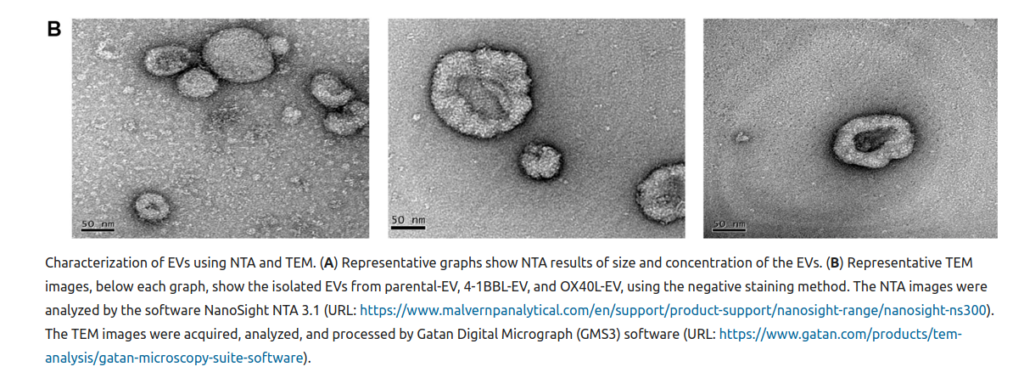
Characterization of extracellular vesicles using electron microscopy

Extracellular vesicles of genetically modified tumor cells may be the key to developing therapies to produce vaccines that can activate the immune system in cancer treatment, as described in a study by CNPEM researchers and published in Scientific Reports.

Marcio Chaim Bajgelman, who led the research team, explains that the goal of developing antitumor vaccines (unlike prophylactic vaccines that can prevent diseases) is to find safe and efficient mechanisms to treat cancer as soon as it is diagnosed.

Since 2017, CNPEM has published studies to develop new skin cancer treatments, with excellent results from using genetically modified tumor cells to express specific combinations of immunomodulators.

The efficacy of activating the immune system via direct contact between modified cells to express immunomodulators on the surface of lymphocytes has already been demonstrated. Animal cell assays demonstrated not only elimination of the tumor, but the capacity to react if the disease recurs.

LYMPHOCYTES CAN RECEIVE ACTIVATION SIGNALS EVEN WHEN NOT IN DIRECT CONTACT WITH ANTITUMOR VACCINES.

The observation that T cells (from the immune system) could also be activated when immersed in the same culture medium in which genetically modified tumor cells had previously been cultured led to a new research phase: this time searching for immunomodulatory proteins that control immunity within this specific medium.

Bajgelman states, “We observed that these proteins we were looking for were actually extracellular vesicles that carried antitumor immunomodulatory proteins.”

In in vitro (in laboratory) testing, extracellular vesicles from modified cells were seen to induce T-cell proliferation and inhibit an immunosuppressant in the regulatory T cells. Since regulatory T cells can inhibit the activity of antitumor lymphocytes, reducing immunosuppressive activity can enhance the effect of antitumor therapies.

Extracellular vesicles are one way cells are known to interact. Since they are naturally produced by all cells (and in especially large numbers by tumor cells), they may offer options to develop new therapeutic approaches to treating cancer in which engineered vesicles carry immunomodulators to stimulate the immune system against tumor cells, as Bajgelman explains:

“Tumor cells use deception so that our immune system tolerates them. Our challenge is to stop this tolerance, making the immune system more effectively recognize the cells that need to be eliminated.”

He adds that although the tests were conducted with melanoma cells, the findings about the role of extracellular vesicles in modulating the immune system open up innovative perspectives for research to develop antitumor vaccines for other types of cancer.

doi.org/10.1038/s41598020-72122-3

Funding Agencies: FAPESP e CAPES

ARTIFICIAL INTELLIGENCE
thisisengineering-raeng-8hgmG03spF4-unsplash

Machines that diagnose

CNPEM USES MACHINE LEARNING IN A NEW DEVICE THAT DIAGNOSES DISEASES WITHOUT REAGENTS OR ANTIBODIES.

In an article published in the journal ACS – American Chemical Society, researchers from CNPEM present a new concept for clinical analysis based on a microfluidic electrochemical sensor and machine learning models that can potentially make the diagnosis and prognosis of several diseases more practical and economical. This method is intended to provide safe results without dependence on expensive and scarce materials (such as antibodies).

WATCH THE PENNIES

The microfluidic device uses low-cost materials: graphite electrodes made from pencil lead (the same kind used by students) act as sensors for electrochemical patterns. When connected to portable equipment that can measure electrical impedance (a potentiostat) and a smartphone, the device can detect the presence and concentration of relevant biomarkers in samples using minimal volumes of blood in less than 15 minutes. “These data not only help screen cases but can also provide references for prognostics of disease progression in each patient,” explains CNPEM researcher and leader of the study, Renato Sousa Lima.

BREAST CANCER

The study describes applying this method to diagnose breast cancer in blood samples taken from mice. Extracellular lipid structures and proteins present in their membranes were used as biomarkers of Ehrlich’s tumor to identify healthy animals and those with this type of cancer. The method also made it possible to simultaneously quantify these two biomarkers, contributing to a high-accuracy analysis of the breast cancer stage. The group prepared the samples in partnership with the USP Institute of Chemistry in São Carlos (IQSC/USP).

MACHINE LEARNING

Each blood sample generates a unique capacitance spectrum that serves as a fingerprint and can generate up to 100 variables. Identifying specific patterns of interest is a task for the machine learning computer model, “a class of methods capable of discovering patterns using only the observed data without being explicitly programmed, which allows it to become even more efficient as new samples are added to the test database,” explains Adalberto Fazzio, researcher and current director of the Ilum School of Science, which was recently inaugurated by CNPEM.

This study also utilized a statistically robust mathematical pattern (algorithm). “Even with a small number of samples (12) for algorithm acquisition, it has demonstrated accuracy and should become even more efficient by incorporating data from more samples in the future,” adds Renato Sousa Lima.

DEVICE FEATURES

SIMPLE AND SCALABLE PRODUCTION

Starting at 1000 units, unit cost is estimated at US$ 0.60

PORTABLE POWER SWITCH FOR ELECTROCHEMICAL MEASUREMENTS

Commercial units can be acquired for US$ 392.90 (Mar 2022), but because their electronics are in the public domain they can be easily constructed at an estimated cost of US$ 98,22 (Mar 2022)

MICROFLUIDIC ELECTROCHEMICAL SENSOR

(US$ 0.60)

SMARTPHONE

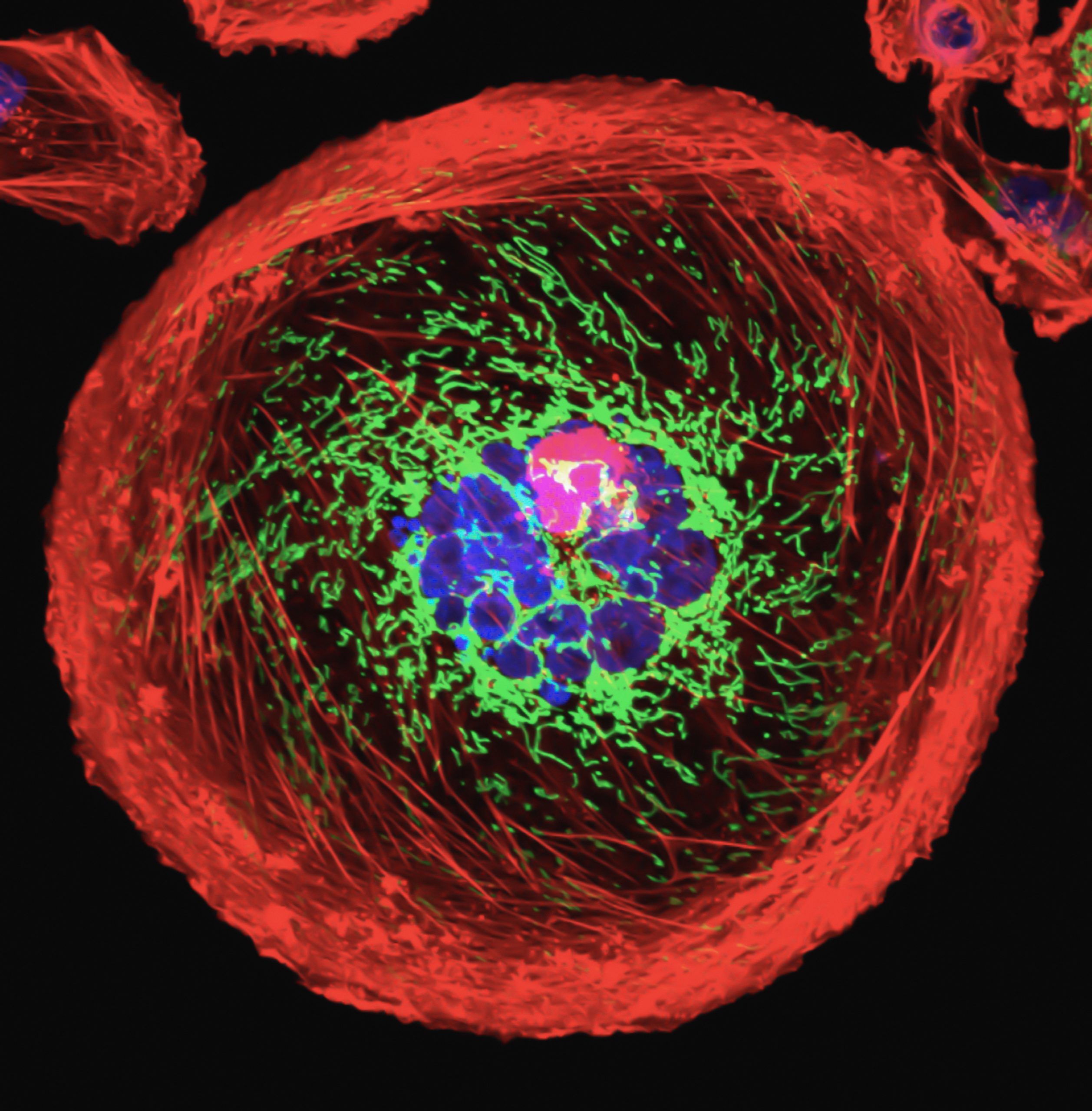
Breast cancer: polyploid giant cancer cell (PGCC)

SIMPLICITY AND TELEMEDICINE

Besides their efforts in science and engineering, the authors utilized technology to combine simplicity and telemedicine with their analyses. A smartphone was used to control a portable device for electrochemical measurements, acquisition and processing of sensor data using machine learning, and finally to present the results of interest on the screen, eliminating any steps involving data processing by the user. In addition to making the analysis simpler, smartphones can be very useful as a support tool for health programs in remote parts of the country. The data obtained from the tests can be quickly shared and contribute to strategic actions such as referring patients to the nearest treatment centers.

TEST PHASES

First, the blood samples must be prepared before being tested by the diagnostic device. The process takes about ten minutes, is simple, and the estimated cost is just under ten Amerian dollars (Mar 2022) per sample. The sensor scan takes less than five minutes and the diagnostic result is automatically displayed on the smartphone screen.

https://pubs.acs.org/doi/10.1021/acssensors.0c00599

Funding Agencies: FAPESP, INCTBio

ENZYMES

Replicating the force of nature, one enzyme after another

INSPIRED BY NATURE, CNPEM RESEARCHERS ARE ATTEMPTING TO UNDERSTAND THE ORGANIZATION OF INVISIBLE STRUCTURES THAT CAN REVOLUTIONIZE GLOBAL MANUFACTURING.
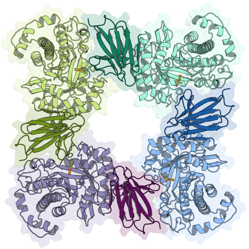
Representação da estrutura
cristalográfica de uma enzima
beta-galactosidase. Pesquisadores
utilizam estratégias de
cristalização e técnicas de
difração de raios X, hoje presentes
nas linhas do Sirius, para obter os
dados e posteriormente visualizar
as estruturas em computador.

Crystallographic structure of a beta-galactosidase enzyme. Researchers use crystallization strategies and X-ray diffraction techniques that are currently part of the Sirius research lines to obtain data and then visualize these structures via computer

Understanding what cannot be seen. Dealing with the unknown. Breaking down concepts into forms and reassembling these forms into biological machines. Uncovering the latent universe of enzymes involves all these stages, and conquering their mysteries requires a lot of science and creativity. Enzymes (which are mostly proteins) are invisible to the naked eye and known to be catalysts: molecules that can accelerate chemical reactions that are essential for the function of complex organic systems such as the human body.

But the body represents only a small sample of enzymatic activities. They are everywhere and used in a multitude of processes from vaccine production to neutralizing lactose in milk for those who cannot tolerate this sugar, from producing biofuels to developing renewable chemicals.

In bioproduct manufacturing, for example, enzymes can be applied to wastes resulting from agricultural processes in large quantities such as residue from orange juice and sugar production, straw, and other materials that until recently were considered low-value-added byproducts. This process known as cleavage makes it possible to obtain acids, solvents, polymers, and advanced biofuels, always using renewable sources and biological approaches.

Enzymes are consequently tools that can completely replace petroleum-based reagents that are currently used to transform molecules into high value-added products. The shift from an oil matrix to biological matrix is equally effective, but with dramatically less environmental impact. The challenge lies in finding the most appropriate enzymes for each desired application.

Identifying these molecules and applying them to the best tasks mobilizes large teams and state-of-the-art instrumentation based on synchrotron light, atomic tools, and bioinformatics. Between 2020 and the first half of 2021, CNPEM researchers identified a group of hitherto unknown enzymes and clarified the mechanism of action for another protein. In the study published in 2020 in Nature Chemical Biology, scientists revealed how a new family of enzymes applied innovative mechanisms and strategies to break down plant polysaccharides and generate useful byproducts such as prebiotics and biofuels.

In 2021, this time in Nature Communications, a new study shattered paradigms with the revelation that enzymes which act on hemicellulose can cleave glycoside connections (converting substrates into products) by two catalytic routes, contrary to prior expectations. In scientific terms, a catalytic route or itinerary represents the chemical and structural changes that a substrate (such as polysaccharides and carbohydrates) undergoes due to the action of the enzyme until it is converted into a product. This set of modifications was considered unique for a given enzyme and its substrate, but the study described pathways that could modify the theoretical understanding.

SEEKING INSPIRATION FROM EVERYTHING AROUND US

Both projects have a common inspiration: nature. Fungi and bacteria are enzyme-producing machines. When a pathogen attacks a plant, for example, the physical barrier is broken by enzymes that disintegrate the plant cell wall and allow the host to enter. Despite the undesirable effects on plants, this is the type of strategy researchers are looking at.

“We observe nature and formulate hypotheses. If an enzyme can rupture the physical barrier in plants, it may be able to do the same with agro-industrial waste, accessing trapped sugars and making them available for fermentation and conversion into products,” says Mario Murakami, scientific director of the LNBR and lead author of the studies.

Absorbing this knowledge and reproducing it in the laboratory is another important front for the Laboratory. CNPEM develops biological platforms (microorganisms) “engineered” via genetic modifications that can be customized to produce enzymes made to order, with potential applications in different activities.

“Our goal is simple: to emulate and improve molecular strategies found in nature that have been shaped over hundreds of millions of years,” he explains.

In CNPEM, enzymes are subjected to crystallography techniques with synchrotron radiation using Sirius, for example, in order to visualize the three-dimensional structure. Other steps include changes in molecular organization, laboratory testing, and pilot plant scaling to determine the feasibility of applying processes in supercritical environments such as manufacturing.

DO ENZYMES WORK ALONE?

The saying “two heads are better than one” can be extrapolated to enzymes. In general terms, different enzymes are synergistically combined to carry out a given activity. For example, dismantling agro-industrial byproducts into fermentable sugars requires a veritable arsenal comprising dozens of enzymes, which we call enzymatic cocktails.

“These enzyme formulations are complex combinations that make it possible to extract components of agro-industrial waste that can be biologically transformed into products of interest to society.”

The CNPEM has experience with such cocktails; one was developed to produce cellulosic ethanol (also known as second-generation ethanol) and patented in 2019 by the National Institute of Industrial Property (INPI) via the Patent Cooperation Treaty (PCT). In this way, CNPEM controls all the stages involved in producing this enzymatic ordnance: engineering the biological platforms (microorganisms that produce enzymes), customizing the enzymes, and developing the cocktail.

These characteristics put Brazil in a prominent position in both scientific and technological terms. By owning the enzyme development and production chain, the country is more competitive, with manufacturers needing to import less and consequently reducing their carbon footprints by using enzymes made locally or even on site.

THE ARTICLES MENTIONED IN THE TEXT ARE ACCESSIBLE AT

doi.org/10.1038/s41589-020-0554-5
Funding Agencies: FAPESP e CNPq

doi.org/10.1038/s41467-020-20620-3
Funding Agencies: FAPESP, CNPq, Spanish Ministry of Science and Innovation (MICINN, AEI/FEDER, UE), Spanish Structures of Excellence María de Maeztu e Agency for Management of University and Research Grants (AGAUR).

NANOTOXICOLOGY

Disinfecting at the nanoscale

BLEACH MAY REDUCE THE TOXICITY OF GRAPHENE OXIDE.

Safe degradation and disposal of nanomaterials is a technological challenge that CNPEM investigates to prevent impacts on human health and the environment.

Graphene oxide is a strategic carbon-based nanomaterial that holds promise for areas of manufacturing that utilize cutting-edge technology: it can alter or add new characteristics such as viscosity, mechanical resistance, and electrical conductivity to products such as paints, filters, packaging, catalysts, electronic devices, biomedical materials, in home construction and many other areas.

Unlike other phases of technological and industrial development, the nanotechnology era involves a global conscience and awareness that incorporation of new materials must also include more attention to waste management, considering the product’s complete life cycle. This starts with the raw material, passes through manufacturing to disposal, and always considers sustainability in the process.

A commitment to health, the environment, and safety is one of the references that guide the nanomaterials research conducted at CNPEM. Along these lines, a recent study has revealed a simple and promising chemical method to degrade and reduce the toxicity of graphene oxide.

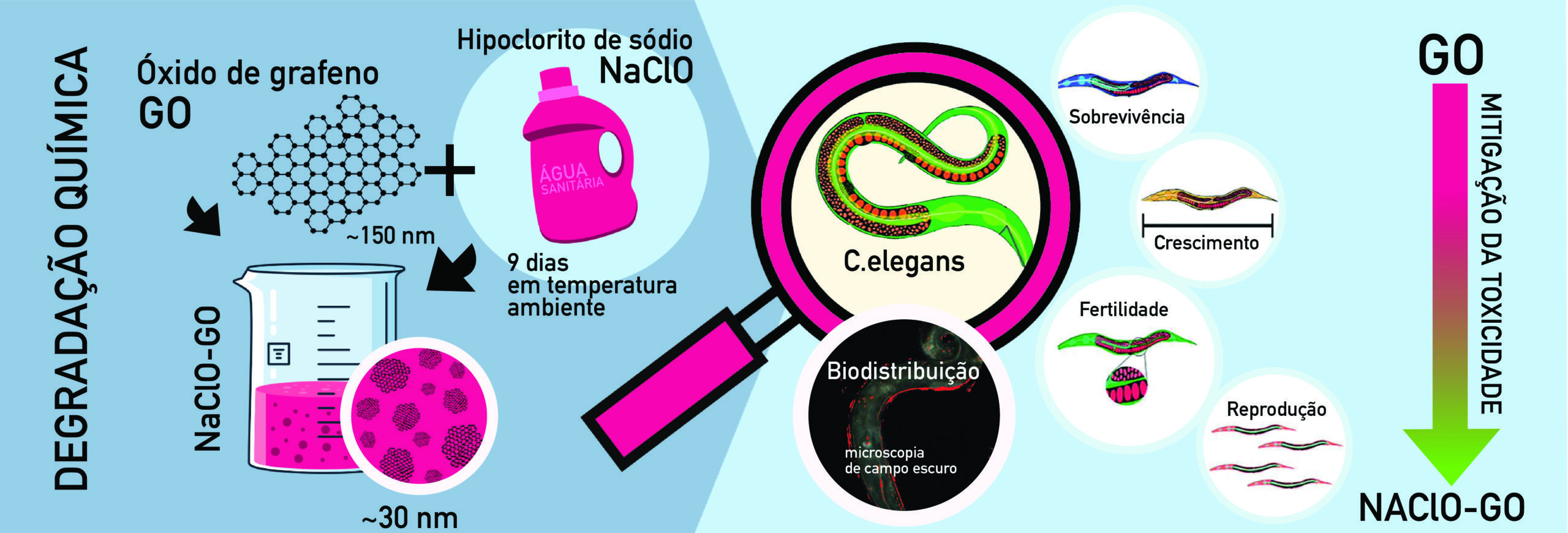
The study, published in Chemosphere magazine, describes results obtained from chemical degradation of graphene nanomaterial using a very common, low-cost product: sodium hypochlorite, better known as bleach.

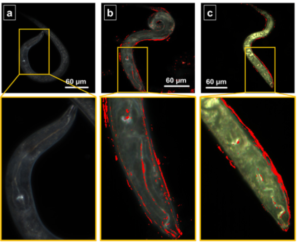
Dark-field hyperspectral microscopy (EDHM) images of the C. elegans nematode model: a) control; b) exposed to graphene oxide; c) exposed to degraded graphene oxide. Red dots indicate the graphene materials adhered to the skin and intestine of the nematode after 24 hours of exposure.

THE STUDY

According to the research, the graphene oxide nanomaterial degrades and becomes much more soluble and less toxic after incubating in bleach for a week.

“The degraded graphene oxide particles shrink to less than 30 nanometers in diameter; [particles] that are not modified or degraded are about 150 nanometers in diameter,” says researcher Diego Martinez.

In many cases, reducing particle size could increase the toxicological risks of graphene, making it more dangerous. Toxicity was tested in Caenorhabditis elegans, a 1-millimeter nematode isolated from soil and commonly used in toxicology testing models. Contact with the graphene oxide nanomaterial affected the survival, growth, and reproduction of these organisms, but this was not observed when they were exposed to the degraded material. Hyperspectral microscopy resources were utilized to confirm the interaction and oral absorption of the nanomaterials; this advanced technique makes it possible to track nanoparticles within biological tissues at high resolution.

“After degradation of the graphene oxide, we observed a reduction of approximately 100% in acute toxicity, as well as a lack of effects on fertility and reproduction in the organisms. So we imagine that this can be a useful method to mitigate risk and safely dispose of graphene-based waste material,” explains Martinez.

NEXT STEPS

This project contributed to a better understanding of transformation of graphene-based materials and their relationship to toxicity (mitigation), which are fundamental to improve management of waste generated by this type of material during its life cycle, but additional studies are necessary to put this methodology into practice.

“Obviously, we will have to test these degraded graphene materials in other animal models, such as bacteria, algae, fish, and human cells. We are already doing this, and we are also studying the interactions with other materials present in waste effluent in order to understand the potential effects on living organisms and environmental impacts in an integrated manner,” adds Diego Martinez.

doi.org/10.1016/j.chemosphere.2021.130421
Funding Agencies: CAPES e INCT-INOMAT

Using nanotechnology against environmental contamination

RESEARCH REVEALS MECHANISMS OF ACTION FOR IRON NANOPARTICLES USED TO DECONTAMINATE AQUIFERS.

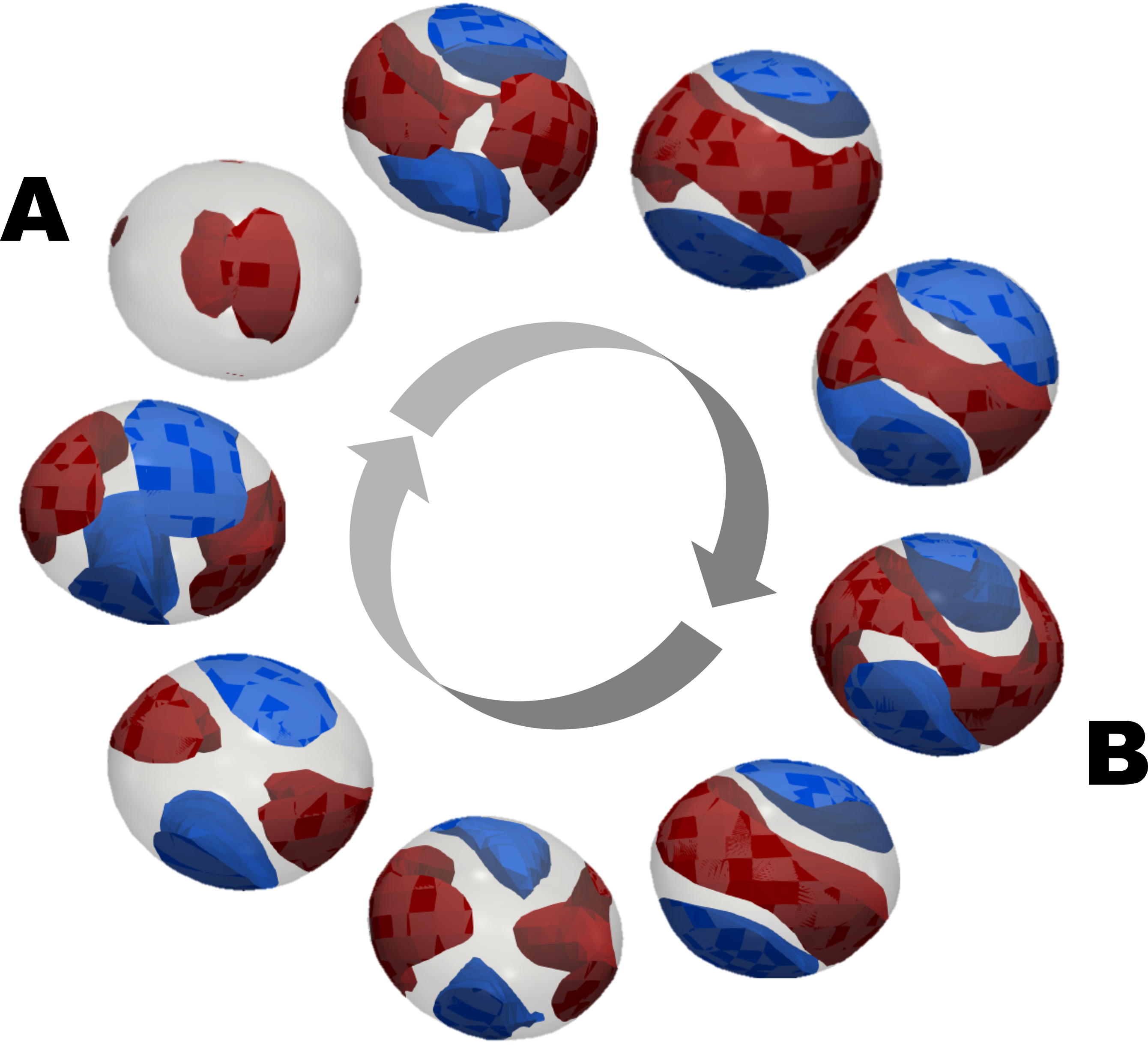
Left to right: gradual degradation of TCE droplets (red) by iron nanoparticles (green) with gas formation (yellow)

Chlorinated hydrocarbons are among the most persistent contaminants in aquifers around the world. This problem is typical of industrialized regions, where these substances were widely used until the 1980s in applications such as solvents, degreasers, and in automotive paint enamel. Very small amounts of these pollutants are enough to make water in these areas unfit for human consumption, since they cause damage to the kidneys and liver as well as cancer.

Because they are denser than water, chlorinated hydrocarbons sink to less permeable regions, usually the bed of an aquifer. These pollutants stay there in the pores of the rocks for many years, and are slowly carried away by the water to regions far from the source of the contamination.

A study published in the journal PNAS describes work by Nathaly Archilha, more CNPEM researchers and colaborators who investigated the use of nanoparticulate iron, a highly reactive chemical element, to degrade one of these pollutants, trichloroethylene (TCE). Although this degradation effect is already known, the group showed for the first time how this reaction occurs in conditions similar to those in a real aquifer, namely what happens in the pores of the rocks during the interaction between the nanoparticles and contaminants.

doi.org/10.1073/pnas.1918683117
Funding Agencies: FAPESP, UK Research and Innovation (UKRI), Teesside University
NANOMATERIALS

Foam made from natural materials found to efficiently absorb oils and solvents

INNOVATIVE COMPOUND OF NATURAL RUBBER LATEX AND CELLULOSE NANOFIBERS FEATURED ON THE COVER OF ACS APPLIED NANO MATERIALS.

Researchers at CNPEM developed a 100% green process to produce a type of foam that can be very useful in decontamination work to remove pollutants from water.

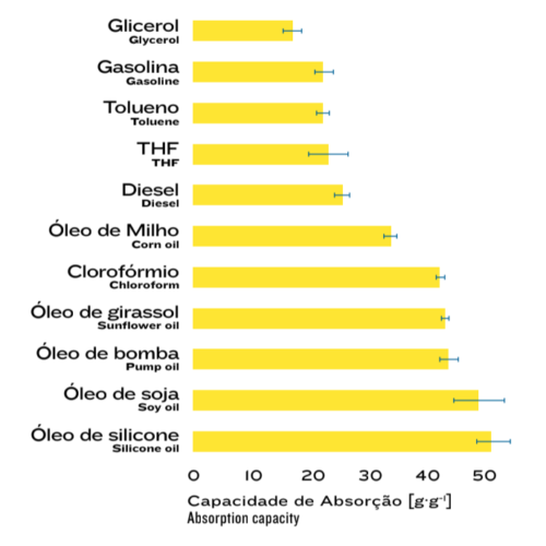
The material, obtained from a combination of nanocellulose fibrils and natural rubber latex, was shown to have an excellent capacity to absorb various types of organic oils and solvents. The scientific article containing the details of the experiments was featured on the cover of the November 2020 issue of ACS Applied Nano Materials.

The process to produce this foam is notable because it uses no petroleum-derived products, only natural materials that are abundant in nature and water as a solvent.

Researcher Rubia Figueredo Gouveia coordinated the study and explains that nanocellulose alone does not have an efficient affinity to absorb the analyzed pollutants, and its fragile structure very easily unravels when it comes into contact with water.

“The combination with natural rubber latex provides the strength needed for the 3D structure of the material and the ability to absorb pollutants.”

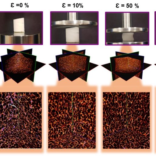
Percentual de compressão da espuma e
imagens das fibras sob pressão.

Percentage of foam compression and images of fibers under pressure

doi.org/10.1021/acsanm.0c02203
Funding Agencies: CNPq

NANOCELLULOSE/NATURAL RUBBER
LATEX FOAM’S CAPACITY TO ABSORB
VARIOUS ORGANIC SOLVENTS AND OILS

Screen Shot 2022-03-14 at 22.46.01

PATENTED EFFICIENCY

This innovation (which CNPEM has the patent) has various possibilities for applications in filters and environmental remediation processes, and has proven ideal characteristics for porosity and structure strength. In addition to the evident differential that comes from using renewable biomass that is abundant from manufacturing, the foam can absorb up to 50 times its mass in pollutants, even those that are more viscous. Tests also showed that the foam retains its high absorption capacity even after 20 re-use cycles, as well as quick absorption (between 1s and 10s).


THE PROCESS IN 3D MICROIMAGES

The foam is obtained in a relatively simple and fast process; the solid compounds (nanocellulose and natural rubber latex) account for only 2% of the composition of the mixture dispersed in water. According to Gouveia, this 98% water is fundamental to obtain the foam’s porous structure, which consolidates after the compound passes through freezing and lyophilization steps.

Scanning electron microscopy and 3D microtomography images obtained from CNPEM laboratories reveal the details that make the difference and demonstrate the reasons behind the efficient affinity between the materials.

“The nanocellulose fibrils are grouped into a 3D structure. After being covered and adhered by the latex, they reorganize into a stronger porous structure that interconnects and contributes to both the robustness and stability of the material and greater absorption of the pollutants,” Gouveia notes.

CATALYSTS

GOLD RUSH

RESEARCH INVESTIGATES THE FORMATION OF ACTIVE SITES IN GOLD CATALYSTS.

Catalysts are substances that promote and accelerate chemical reactions without being consumed in the process. They are widely used not only in industrial manufacturing processes, but also to control pollutant emissions, for example from motor vehicle exhaust, where catalytic converters are used to reduce emissions of toxic gases such as carbon monoxide.

In catalysts composed of nanoparticles of transition metals, the catalytic reaction occurs only in specific places on the surface known as active sites, which are usually located in superficial defects such as regions of deformation in the distribution of atoms that comprise the nanoparticles.

Understanding the dynamics of the formation and evolution of active sites during reactions is crucial to develop economically viable, efficient, and stable catalysts. For this reason, CNPEM researchers and their collaborators investigated the changes in deformation of the atomic network of gold catalysts during the oxidation reaction of carbon monoxide (a reaction similar to that used in vehicle exhaust), in a study published in Nature Communications. They observed the dynamics of nanoscale deformation, and how deformation leads to the formation of active sites, enabling the search for new ways to control the catalytic properties of nanomaterials.

doi.org/10.1038/s41467-020-18622-2
Funding Agencies: FAPESP

Florian Meneau and the research group observed the dynamics of deformation at the nanometer scale, and how the deformation leads to the formation of active sites, allowing the search for new ways to control the catalytic properties of nanomaterials

BACTERIA

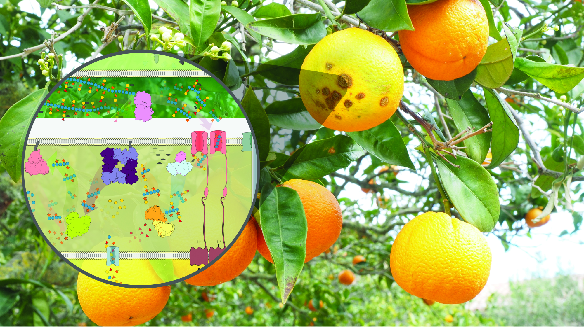
Citrus pest proves to be an ally in the production of biorenewables

VILLAIN OR HERO?

The bacterium Xanthomonas citri, responsible for citrus canker, has always been known as the enemy of the crop because of the damage it causes to citrus farming. But the work led by CNPEM and published in the journal Nature Communications, in collaboration with USP and Unicamp, reveals valuable details about the depolymerization mechanisms of complex vegetable carbohydrates, for the development of new technologies that can be applied in the use of agro-industrial residues, contributing for a more sustainable economy. The study showed that the same biological processes used by the bacterium to weaken one of the most important defence systems of plants, the plant cell wall, can serve as the basis for the development of enzymatic technologies, that enable the use of plant biomass as a raw material for the production of biofuels, biochemicals and biomaterials, serving as an alternative for the production of various products that are still obtained almost exclusively from petroleum derivatives today.

THE PATH OF DISCOVERY

Over five years, a wide range of scientific resources available in CNPEM research ecosystem was used, such as synchrotron beamlines, genomics, transcriptomics and genetic engineering approaches and in vivo experiments in plants to complete the work. The research uncovered a hitherto unknown family of enzymes that the bacterium X. citri mobilizes to deconstruct the cell wall of plants. This new family of enzymes was named CE20, an acronym for Carbohydrate Esterase Family 20. In addition, this research also revealed the importance of depolymerization of one of the most complex carbohydrates in the plant cell wall, xyloglycan, for the release of specific sugars that induce the production of proteins that potentiate the infection. The efficiency of the work of enzymes is due to their exceptional ability to act on chemical bonds and degrade complex carbohydrates.

“These discoveries provide us with new alternatives to increase the capacity to use plant biomass in biorefineries, which are very valuable from a biotechnological point of view. And by revealing new components of the virulence of the bacterium, we can collaborate with the development of new strategies to combat the disease, with the design of potential inhibitors for this group of bacteria so relevant to Brazilian agriculture”, explains Mario Murakami, research coordinator and scientific director of the Brazilian Biorenewables National Laboratory (LNBR).

NEXT STEPS WITH SIRIUS

On Sirius, research goes to a new level. Let’s move beyond static images, such as photos, to start seeing and analyzing dynamic events, as in videos of the catalytic processes of the enzymes discovered in this study”, exemplifies Murakami.

Combining the results obtained from the MANACÁ beamline from Sirius with quantum calculations will provide atomic details of all steps of a catalytic reaction. CNPEM has developed several studies that have been internationally recognized for their impact on the development of processes that can accelerate the industrial production of bioproducts for various economic segments. More sophisticated biochemicals and materials, with high performance and efficiency, greater added value and less environmental impact are the focus of the search for more sustainable alternatives to fossil-based inputs used in the industry today.

INFRARED

Capturing light in nanobelts

RESEARCH INVESTIGATES CONFINING LONG INFRARED WAVES IN TIN OXIDE NANOBELTS.

Long infrared is low-energy, non-destructive radiation suitable for applications in biological materials. It also penetrates materials to a high degree, which makes it useful in non-invasive inspection of goods and people.

Infrared nanophotonics has investigated new materials (such as graphene) to explore their properties and use in this energy range. Within this context, nanostructured semiconductor oxides have gained relevance given the variety of forms in which they can be synthesized, which include nanoparticles (0D), nanobelts (1D), nanosheets (2D), and nanocubes (3D).

Researchers from CNPEM and collaborators from Brazil and abroad studied the confinement of long infrared waves in tin oxide (SnO2) nanobelts in a study published in Nature Communications. Ingrid Barcelos and the group found that SnO2 nanobelts offer an excellent nanophotonic platform to confine long infrared waves, and are naturally optimized for applications in components and circuits for new information networks based on light traffic.

Infrared nanophotonics has been dedicated to studying new materials, such as graphene, in order to explore their properties and use in this energy range

doi.org/10.1038/s41467-021-22209-w
Funding Agencies: INCT/Nanocarbono, CNPq, FAPESP, FAPEMIG, BMBF

USER PUBLICATIONS

Although there has been little in-person movement at the CNPEM to use the open facilities over the past two years, researchers have continued their research and collaborations. Articles involving data collected at the open facilities in the four national laboratories were published in periodicals in the northern and southern hemispheres, and contributed to global scientific production in various knowledge areas. Below are some highlights from 2020 and 2021.

MICROSCOPY

Visualizing wear in the welding process

Researchers from USP, Unicamp, UNESP, SENAI, the CNPEM, the National Institute of Technology, Petrobras, and The Ohio State University used LNNano’s electronic microscopy equipment for research that led to an article entitled “The study of volumetric wearing of PCBN/W-Re composite tool during friction stir processing of pipeline steels (X70) plates,” which was published in 2021 in the International Journal of Advanced Manufacturing Technology, and funded by Petrobras.

The study analyzed the friction stir welding technique for steel, under controlled conditions. The wearing that occurs during this process was observed via profilometry and light microscopy to quantify the loss of volume in the welding tool resulting from the number of plunges and the welded distance. Torque and transverse force on the welding tool and the width of the weld bead were measured and related to the wear process.

doi.org/10.1007/s00170-021-06932-8
Funding Agencies: Petrobras
THE ENVIRONMENT

Lasting consequences of the Mariana disaster

The Fundão dam breach in the town of Mariana, Minas Gerais on November 5, 2015 is considered the largest environmental disaster in Brazilian mining industry. The resulting flood of over 40 million cubic meters of iron ore tailings caused the death of 19 people, total and partial destruction of villages, as well as untold environmental damage. After the tailings swept through the Doce River basin, some reached the mouth of the Doce in Linhares, Espírito Santo, and the adjacent region in the Atlantic Ocean.

The composition of mining waste depends on the rocks processed, chemicals used to extract the iron ore, efficiency of the process, and breakdown of the tailings during storage in the dam. This composition serves as a fingerprint to differentiate between material that came from the dam and other materials that were already present in the marine sediment, whether natural or of other anthropogenic origin.

In an article published in the journal Chemosphere, researchers from the Federal University of Espírito Santo (UFES) and their collaborators used CNPEM’s UVX synchrotron light source to study the metals present in tailings from the Fundão dam and in the marine sediment adjacent to the mouth of the Doce River before and after the accident. The group found that the metals introduced into the ocean floor from the tailings had not yet returned to pre-disaster levels, and that it is not yet possible to predict how long the tailings will persist in the marine environment.

doi.org/10.1016/j.chemosphere.2020.127184
Funding Agencies: CAPES, FAPES
METABOLISM

Does exercise without dieting work?

Physical training and healthy eating are the main non-pharmacological strategies for treating chronic conditions such as obesity and insulin resistance in adolescents. But isolated metabolic changes resulting from physical training without dietary interventions have not yet been established. Researchers from Unicamp, in collaboration with the State University of Maringá and the Federal University of Paraná and with funding from CAPES and CNPq, published an article in Scientific Reports in October 2020 entitled “Altered metabolomic profiling of overweight and obese adolescents after combined training is associated with reduced insulin resistance,” in which they analyzed the metabolic characteristics of 37 overweight and obese adolescents after they completed 12 weeks of physical training without consuming specific diets. The researchers used LNBio’s nuclear magnetic resonance facility to obtain data, and observed positive effects from training on the metabolomic profile, body composition, biochemical markers, and glucose metabolism of the study participants.

doi.org/10.1038/s41598-020-73943-y
Funding Agencies: CNPq e CAPES
FUNGUS

Mechanisms of fungal propagation

The article “The Aspergillus fumigatus transcription factor RglT is important for gliotoxin biosynthesis and self-protection, and virulence,” published in July 2020 in PLOS Pathogens, was the fruit of cooperation by researchers from Brazil, China, the United States, England, and Ireland, from USP, the Butantan Institute, UNIFESP, the University of Macau, Vanderbilt University, the University of Manchester, and Maynooth University, respectively.

The study defined important mechanisms of the fungus Aspergillus fumigatus, an opportunistic pathogen that causes several mammalian diseases and secretes an immunomodulatory and immunosuppressive mycotoxin (a fungal toxin) that is significant for its virulence.

The researchers used the LNBR’s DNA sequencing facility and found that a transcription factor known as RglT is essential as the fungus protects itself and synthesizes gliotoxin, a process that mainly occurs via direct regulation of one gene; without this gene, the fungus becomes highly sensitive to oxidative stress. The researchers also observed this mechanism in non-pathogenic fungi, and concluded that self-protection via mycotoxins in pathogenic and non-pathogenic fungal species is important to their virulence and survival.

doi.org/10.1371/journal.ppat.1008645
Funding Agencies: FAPESP, CNPq, CAPES, National Science Foundation, Vanderbilt University,
James H. Gilliam Fellowships for Advanced Study program, Wellcome Trust, University of Macau, Institute of Translational Medicine and Faculty of Health Sciences internal funds